Introduction
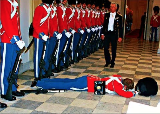
Syncope is an abrupt, transient, spontaneously-resolving loss of consciousness and postural tone caused by a transient drop in cerebral perfusion. It is a descriptive and an interim diagnosis which requires further clarification.
The first step
Syncope should be distinguished from alternative causes of loss of consciousness and postural tone. Syncope mimics include seizures, concussions, sleep (both normal or narcoleptic), migraines and various psychogenic conditions. History from a witness is almost always the best initial diagnostic step. In patients with syncope, both the prodrome and aftereffects are fairly benign (unless the patient crashed a vehicle in the process…).
The next step is to evaluate the patient for possible red flags:
- The worrisome history of present illness: The following cluster of findings almost always points to a worrisome cause: absence of a prodrome, passing out with an appearance (to witnesses) as if the patient was lifeless, a sudden and prompt restoration of consciousness and of all mental faculties, all in the absence of psychogenic, vasovagal or orthostatic provocation.
- Chest pain → myocardial ischemia or infarction
- Shortness of breath → pulmonary embolism
- Headache → subarachnoid hemorrhage
- Abdominal or back pain → abdominal aortic aneurism
- Exertional syncope → either an obstructive cause (such as aortic stenosis) or an effort-induced arrhythmia (commonly ventricular tachycardia). Therefore, these patients should get an echocardiogram and possibly an exercise electrocardiogram.
- The worrisome past medical history: History of structural heart disease, particularly of ischemic cardiomyopathy and valvular heart diseases (especially aortic stenosis).
- The worrisome family history: family history of syncope or of sudden cardiac death at a young age points to something bad, namely hypertrophic obstructive cardiomyopathy (HOCM), long QT syndrome, or Brugada syndrome. The younger the family member was at time of sudden cardiac death or syncope, the redder the red flag.

An electrocardiogram, when done, should be scrutinized for:
- T-wave inversions:
- Ischemia (new or old): look for ST changes, Q waves (especially new or worsening), or T wave inversions.
- Giant T-wave inversions: sometimes seen (particularly in precordial leads) in patients with subarachnoid hemorrhage → CT the brain without contrast.
- Brugada Sign: Look for typical or atypical right bundle branch block pattern in leads V1-V3 with a downsloping, coved, elevated ST segment which begins at the top of R’ and ends with an inverted T wave, and without reciprocal changes (Wang, 2013). Before diagnosis Brugada syndrome, make sure that the patient is not hyperkalemic, as hyperkalemia can cause a Brugada sign. Check serum potassium level and look for tall, narrow, pointed and tented T-waves “as if pinched from above.”
- Epsilon waves: these, along with T-wave inversions, are seen in V1-V3 in patients with arrhythmogenic right ventricular cardiomyopathy → confirm with cardiac MRI.
- Dysrhythmias (e.g., bradycardia, which can be suggestive of sick sinus syndrome)
- Conduction abnormalities (especially third degree heart block or Mobitz Type II)
- Evidence of right ventricular strain/pulmonary hypertension/pulmonary embolism:
- sinus tachycardia,
- S1Q3T3, new right bundle branch block or incomplete right bundle branch block,
- T wave inversions, especially in anteroseptal and inferior leads.
- Regardless, “the most important thing to remember about pulmonary emboli is that most of them have no effect on the electrocardiogram (ECG) at all…. The reason is obvious: the lungs have such an abundant blood supply that an embolus must compromise a really major segment of the lung before it has any effect on pressure in the pulmonary artery.” (Phibbs, p. 240)
- Abnormal intervals
- short QT
- Long QTc → ischemia, hypokalemia, hypocalcemia, drugs, congenital
- Preexcitation (Wolff-Parkinson-White) pattern:
- Without atrial fibrillation: look for (1) short PR intervals, (2) Delta waves and (3) wide QRS complexes. Also seen are ST changes and T wave inversions that are discordant with the delta waves.
- With atrial fibrillation:
- An irregular polymorphic tachycardia, with rates as high as 250-300 beats per minute, but only in only some places,
- Changing morphologies, including changes in amplitudes and width, with sometimes deformed and bizarre QRS complexes,
- Look for Delta waves, and pauses as long as two R-R intervals.
- Low voltage → pericardial effusion or tamponade. Look for electrical alternans and sinus tachycardia (or sinus bradycardia → hypothyroidism).
- High left ventricular voltage: in the setting of syncope, high left ventricular voltage suggests either aortic stenosis or hypertrophic obstructive cardiomyopathy (HOCM). In HOCM, the resting ECG is usually nonspecific and ranges from completely normal (up to 25%) to various degrees of high left ventricular voltage, repolarization abnormalities, and left atrial enlargement. Dramatically high left ventricular voltage with deep narrow Q-waves in inferior and lateral leads (I, aVL, V5, V6) is rather specific for HOCM.
- Evidence of pacemaker malfunction. Look for
- pacer spikes that are not followed by P wave/QRS complex (failure to capture),
- pacemaker rate
- spontaneous complexes that do not inhibit the pacemaker
- failure of pacemaker to ‘”take over” when spontaneous rhythm slows down.
Patients with a pacemaker should get a chest radiograph (for lead placement) and a pacemaker interrogation.
Longer range options for rhythm analysis include:
- Telemetry
- Holter monitor: continuous monitoring for several days. Best for frequent events
- Event recorder: activated by patient. Best for infrequent events with a prodrome
- External loop recorder: records continuously in a loop, and a recorded loop is saved retroactively after the event starts. Best for infrequent events without a prodrome
- Implantable loop recorder: records for years. Best for very rare events.
- Exercise electrocardiogram: for exercise-induced dysrhythmias
Key associations:
- Upon sitting or standing up → orthostatic hypotension
- Coughing, urination, defecation → neurocardiogenic
- After a meal → postprandial syncope
- Preceded by palpitations → sick sinus syndrome, arrhythmias
- While shaving, turning head or other forms of carotid sinus manipulation → carotid sinus syndrome
- Vertigo, diplopia, dysarthria, numbness or weakness (neurological prodrome) → vertebrobasilar transient ischemic attack.
- upper extremity exertion and neurological prodrome → subclavian steal syndrome
- Shortness of breath, hypoxia, hemodynamic instability and/or evidence of right ventricular strain on echocardiogram or EKG → pulmonary embolism
- Plethoric facies → superior vena cava syndrome
- Positional syncope or murmur, embolic phenomena → atrial myxoma
- Breath holding in an infant toddler → breath holding spells
References:
- Henderson, Marc C., MD, The Patient History: an Evidence-Based Approach to Differential Diagnosis, 2e (2012)
- Sabatine, Marc S., MD, MPH, Pocket Medicine: The Massachusetts General Hospital Handbook of Internal Medicine, 5e (2013)
- Wang, K., MD, Atlas of Electrocardiography (2013)
- Phibbs, Brendan, MD, Advanced ECG: Boards and Beyond, 2e (2005)
- Pediatric EM Morsels.
- Davey, Patrick, ECG at a Glance (2008)
- Mattu, Amal, MD, Emergency ECG Video of the Week


Leave a Reply