Etiology
- Traumatic: exercise, Foley catheterization, nephrolithiasis, coitus
- Infectious: urinary tract infections
- Malignant: bladder, kidney, prostate
- Vascular: renal infarct, renal vein thrombosis, sickle cell disease and trait
- Toxic: cyclophosphamide, antiplatelet agents, anticoagulants, NSAIDS
- Spurious: menses
Workup
Step 1: discontinue offenders
- Anticoagulants and antiplatelet agents
- Hemorrhagic cystitis: isophosphamide, cyclophosphamide, radiation therapy, methotrexate
- stone forming agents: loop diuretics, anticonvulsants, indinavir
- Renal papillary necrosis: NSAIDS
- Other: blood transfusions
Step 2: look at urine color, perform a urine dipstick, and sedimentary microscopy:
- If dipstick is positive for blood, leukocyte esterase, white blood cells, nitrates, and bacteria, treat for urinary tract infection
- If urine sediment is positive for dysmorphic red blood cells, red blood cell casts, protein, then work the patient up for glomerulonephritis and/or consult a nephrologist
- If urine microscopy is persistently positive for nondysmorphic red blood cells:
- in non-pregnant adults without flank pain, order CT abdomen and pelvis without and with contrast (without to look for stones, with to look for tumors and vascular lesions); in patients with clots or with risk factors for bladder cancer (age, smoking, aniline dyes, schistosomiasis) order cystoscopy and cytologic analysis of urine (to look for bladder tumors that are too small to be seen on CT)
- in patients with acute onset of flank pain that is suspicious for nephrolithiasis, order only CT abdomen and pelvis without contrast (then, only if negative, proceed to CT with contrast to look for non-stone causes such as tumor or renal infarction; an elevated LDH points toward renal infarction)
- in pregnant and pediatric patients, order an ultrasound of the urinary system instead of a CT
- in a trauma patients with gross hematuria, get CT of the abdomen and pelvis without contrast, plus a CT cystogram
- in a trauma patient with blood at the urethral meatus and inability to void, get a retrograde urethrogram and urology consult; do not attempt to insert a Foley catheter
- in a patient with a positive family history and bilateral flank masses, order a renal ultrasound (to look for autosomal dominant polycystic kidney disease)
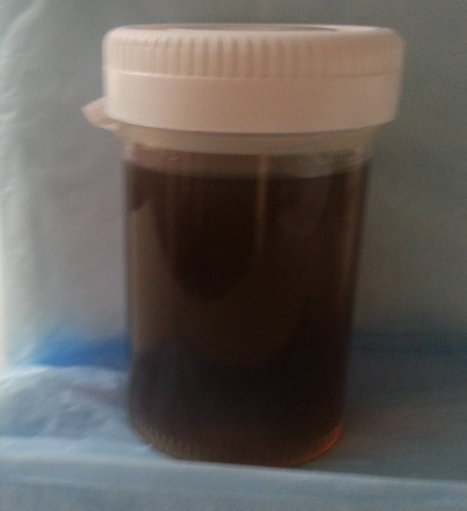
- If dipstick is positive for “blood,” but sediment is negative for red blood cell, then the diagnosis is myoglobinuria or hemoglobinuria. Work the patient up for intravascular hemolysis (CBC, peripheral blood smear, LDH, bilirubin, reticulocyte count, haptoglobin) and for rhabdomyolysis (creatine kinase, urine myoglobin). Note that pink urine points to hemoglobinuria, not to myoglobinuria. If hemolysis and rhabdomyolysis have been ruled out, consider one of the porphyrias
- If the urine looks red, but the dipstick and microscopy are both negative, consider ingestion of beets, blackberries, or rhubarb as possible causes. Orange urine is sometimes seen with rifampin and phenazopyridine (Pyridium)
Step 3: consider workup for systemic diseases in appropriate settings
- Platelet count, PT, PTT, plasma fibrinogen, plasma d-dimer (for suspected disseminated intravascular coagulation)
- Hemoglobin electrophoresis (for suspected sickle cell disease or trait)

Important associations
- irritative voiding: bladder infection, prostatitis, urethritis, bladder calculi, hemorrhagic cystitis,
- flank pain: ureterolithiasis, renal artery thrombosis, renal vein thrombosis, renal cell carcinoma
- unilateral flank mass: renal cell carcinoma
- bilateral flank masses: autosomal dominant polycystic kidney disease
- edema, hypertension, dysmorphic red blood cells: glomerular disorders
- preceded 1-3 weeks by group A β-hemolytic streptococcal pharyngitis or impetigo and presenting with dark urine, proteinuria, renal insufficiency: postinfectious glomerulonephritis (order antistreptolysin-O, anti-DNase B antibodies antihyaluronidase, C3)
- preceded 1-3 days by upper respiratory or diarrheal illness and presenting with hematuria: IgA nephropathy (consider renal biopsy)
- Sinus issues: granulomatosis with polyangiitis (“Wegener’s granulomatosis”)
- Lung issues: Goodpasture syndrome
- Child with:
- abdominal pain, arthralgias, and purpuric, popular rash on buttocks and legs in the setting of a recent upper respiratory tract infection: Henoch-Schönlein purpura; think about intussusception
- hemolytic anemia, thrombocytopenia and acute renal failure after or with a diarrheal illness secondary to consumption of poorly cooked meat: hemolytic-uremic syndrome
- abdominal mass, hypertension and hematuria: Wilms’ tumor; look for aniridia and hemihypertrophy
- With episodic hemolysis, hematuria, with venous thromboembolism: paroxysmal nocturnal hemoglobinuria (order flow cytometry for diagnosis)
- Autosomal dominant inheritance pattern, cerebral aneurism: autosomal dominant polycystic kidney disease
- With weight loss, polycythemia, flank mass: renal cell carcinoma
- In men with incomplete voiding, weak urinary stream: benign prostatic hyperplasia
- Coinciding with menses: spurious vs. endometriosis (catamenial hematuria)
- Smoking: bladder cancer
- Clots: non-glomerular sources
- Abnormal hemoglobin electrophoresis: sickle cell disease or trait
- Travel to Africa: schistosomiasis, malaria (“blackwater fever”)
References
- The Merck Manual for Health Care Professionals
- Buttaravoli, Philip, MD. Minor Emergencies (2012, previous edition reviewed here)
- O’Connell, Theodore X., MD. Instant Workups (2008, reviewed here)
- Sabatine, Marc S., MD MPH. Pocket Medicine (2011)
- American College of Physicians (2012), Board Basics 3 (reviewed here)


Leave a Reply