blood clots or tumours in the brain deep to the pia mater are called intraaxial and those outside the pia are called extra axial.
(P. 26-27)
perhaps the most important clinical advice to the front line doctor with regards to emergency brain CT scans for trauma or any other reason is for you to look at the scan as soon as it is done.(P. 33)
This is straightforward advice and information that, for whatever reason, seldom makes its way into other radiology books.

In other places, the book is much more subtle and sophisticated. According to the author, for example:
The first important clue to early hydrocephalus is enlargement of the temporal horns….
The next important clue … is that the third ventricle which normally presents a narrow slit lie appearance changes to an oval shape.(P. 74-75)
Overall, the reader is left with the impression that the author really, really, knows what he is talking about.
The book disproportionately focuses on subarachnoid hemorrhage and hydrocephalus. This is a very smart decisions because these are critically important diagnoses that can be very difficult to detect without good training. In contrast, hemorrhagic strokes are generally far from subtle in appearance, while ischemic strokes are essentially clinical diagnoses whose management seldom depends upon the findings on head CT.
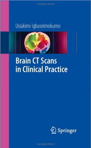
On the downside, the pictures aren’t excellent. They are, however, they are good enough to help elucidate the text which, in turn, is what makes to book exceptionally good.
I recommend this book very highly to residents in primary care and to medical student with a strong interest in internal medicine, emergency medicine, radiology, neurology or neurosurgery. It is one of the core books in Neurology: A Curriculum for Self-Guided Learners and is easily one of the best medical books of all time.
Free sample here.


Leave a Reply